diabetic foot examination pdf

A diabetic foot examination is critical for early detection of complications‚ ensuring timely intervention to prevent severe outcomes. It involves a comprehensive assessment of foot health‚ focusing on identifying risk factors‚ detecting early signs of neuropathy‚ and evaluating vascular integrity. Regular exams are essential for individuals with diabetes‚ as they help in preventing ulcerations and amputations. The International Working Group on the Diabetic Foot (IWGDF) guidelines emphasize the importance of structured foot evaluations to improve patient outcomes and quality of life.
1.1 Importance of Regular Foot Exams for Diabetic Patients
Regular foot exams are vital for diabetic patients to detect early signs of complications‚ such as neuropathy or poor circulation. Early detection prevents severe outcomes like ulcers or amputations. The International Working Group on the Diabetic Foot (IWGDF) recommends frequent assessments to identify risks and ensure timely intervention‚ improving overall foot health and patient outcomes.
1.2 Brief Overview of Diabetic Foot Complications
Diabetic foot complications include ulcers‚ infections‚ and neuropathy‚ often due to high blood sugar and poor circulation. Untreated‚ these can lead to severe infections and amputations. Regular exams help identify risks early‚ while guidelines like those from IWGDF emphasize prevention and timely treatment to manage these complications effectively.
Preparation for the Diabetic Foot Examination
Preparation involves removing shoes and socks for a thorough inspection‚ ensuring proper lighting‚ and positioning the patient comfortably to facilitate an accurate and effective examination process.
2.1 Removing Shoes and Socks for a Thorough Inspection
Removing shoes and socks is essential for a comprehensive foot examination. This allows healthcare providers to inspect all areas‚ including the soles‚ toes‚ and heel‚ for signs of ulcers‚ redness‚ or swelling. Proper removal ensures no areas are missed‚ enabling an accurate assessment of foot health and early detection of potential complications. This step is crucial for effective diabetic foot care.
2.2 Ensuring Proper Lighting and Comfort for the Patient
Proper lighting and patient comfort are vital for an accurate diabetic foot examination. A well-lit‚ private space allows for a detailed inspection of foot anatomy. Patients should be seated comfortably‚ with feet resting flat on the floor or a sturdy surface. This setup ensures optimal visibility and minimizes patient anxiety‚ facilitating a thorough and effective assessment of foot health.
Visual Inspection of the Feet
Visual inspection is a crucial step in diabetic foot exams‚ enabling early detection of potential issues. Healthcare providers examine the feet for any abnormalities.
3.1 Examining the Soles and Between the Toes
During the visual inspection‚ carefully examine the soles and interdigital spaces for signs of ulcers‚ calluses‚ or fissures. Ensure proper lighting to detect subtle changes. Patients should be encouraged to self-inspect these areas regularly‚ using mirrors or seeking assistance if needed. The use of instructional videos or guides can aid in identifying abnormal findings early. This step is vital for preventing complications and ensuring timely intervention. Regular checks can significantly improve outcomes for diabetic patients.
3.2 Checking for Signs of Ulcers‚ Redness‚ or Swelling
Examine the feet for ulcers‚ redness‚ or swelling‚ which may indicate early signs of complications. Pay special attention to pressure points and between toes. Use proper lighting to detect subtle color changes. Patients should be educated to monitor these areas daily‚ using mirrors if necessary. Early identification of such signs is crucial for preventing infections and promoting timely treatment‚ as outlined in diabetic foot care guidelines.
Assessing Foot Sensation
Assessing foot sensation is crucial for detecting neuropathy. Techniques include monofilament testing and evaluating vibration sensitivity to identify nerve damage early.
4.1 Using a Monofilament Test for Peripheral Neuropathy
The monofilament test is a simple‚ effective method to assess peripheral neuropathy in diabetic patients. A standardized 10-gram filament is applied to specific points on the foot. The patient’s ability to feel the pressure indicates nerve function. Reduced sensation may signal neuropathy‚ necessitating further evaluation and preventive measures to avoid complications like ulcers.
4.2 Evaluating Vibration and Pinprick Sensitivity
Evaluating vibration and pinprick sensitivity helps assess sensory function in diabetic patients. A tuning fork is commonly used to test vibration perception‚ while a sterile needle or monofilament applies gentle pressure for pinprick sensitivity. These tests detect nerve damage early‚ enabling timely interventions to prevent complications like ulcers. They complement other assessments in a comprehensive foot exam‚ aligning with IWGDF guidelines for optimal patient care.

Evaluating Foot Shape and Deformities
Evaluating foot shape and deformities involves checking for common structural abnormalities like hammertoes or bunions‚ which can significantly increase the risk of ulceration in diabetic patients.
5.1 Identifying High-Risk Areas for Ulceration
High-risk areas for ulceration include regions with abnormal pressure distribution‚ such as the ball of the foot‚ heels‚ and between the toes. These areas are prone to friction and shear forces‚ especially in individuals with deformities or improper footwear. Early detection through visual inspection and pressure assessment can help prevent complications.
5.2 Assessing for Conditions Like Hammertoes or Bunions
Hammertoes and bunions are common deformities that alter foot structure‚ increasing pressure on specific areas. These conditions can lead to calluses and corns‚ which may progress to ulcers if left untreated. Diabetic patients with such deformities are at higher risk for poor wound healing and infection. Early detection and proper management‚ such as orthotics or shoe modifications‚ are crucial for preventing complications.
Checking for Pain or Numbness
Discuss any history of pain or discomfort with the patient. Perform a systematic assessment to identify areas of tenderness or numbness‚ which may indicate neuropathy or underlying issues.
6.1 Patient History of Pain or Discomfort
Begin by discussing the patient’s history of pain or discomfort in their feet. Ask about the duration‚ location‚ and characteristics of the pain. Inquire if they experience relief or exacerbation with activity or rest. Note any recent trauma or changes in sensation. This information helps correlate symptoms with physical findings and guides further examination. Documenting pain history is crucial for diagnosing neuropathy or underlying conditions.
6.2 Correlating Symptoms with Physical Findings
Correlating symptoms with physical findings is crucial for accurate diagnosis. Physical signs like ulcers‚ redness‚ or swelling often align with patient-reported pain or discomfort. This correlation aids in identifying conditions such as peripheral neuropathy or poor circulation. Documenting these findings ensures comprehensive care and guides treatment decisions. Accurate correlation enhances diagnostic precision and improves patient outcomes in diabetic foot management.
Vascular Assessment
Vascular assessment evaluates blood flow to identify circulatory issues. Techniques like ABI measurement and Doppler studies help detect conditions such as peripheral artery disease‚ ensuring timely intervention.
7.1 Measuring Ankle-Brachial Index (ABI)
Measuring the Ankle-Brachial Index (ABI) is a non-invasive method to assess peripheral artery disease. It involves comparing blood pressure readings from the ankle and arm. An ABI value below 0.9 indicates potential arterial blockage. This test is crucial in diabetic foot examinations to identify patients at risk of poor circulation‚ guiding further vascular evaluation and management to prevent complications.
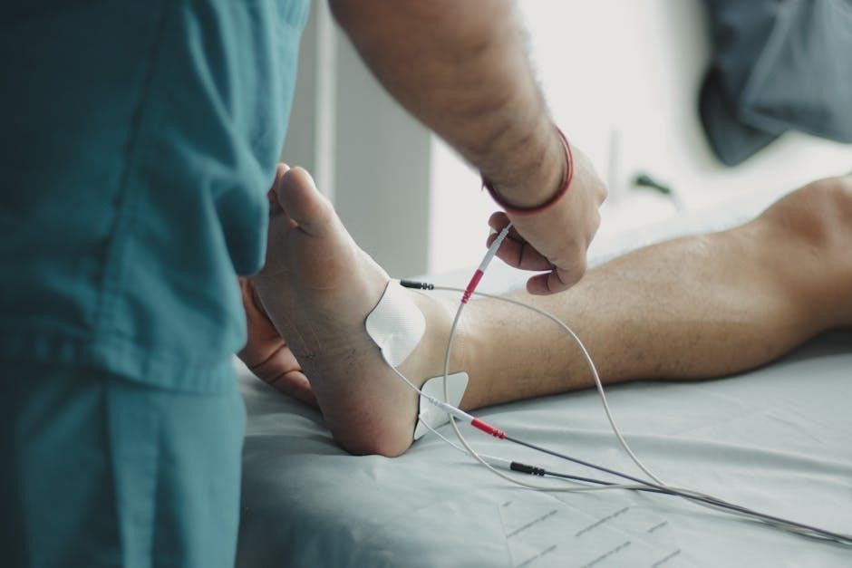
7.2 Performing Doppler Studies for Blood Flow
Doppler studies are essential for evaluating blood flow in diabetic patients. Using ultrasound technology‚ they detect arterial stenosis or occlusion. This non-invasive method provides detailed information about circulation‚ aiding in early detection of peripheral artery disease. Abnormal results may indicate impaired blood supply‚ necessitating further investigation or intervention to prevent complications like ulceration or gangrene.
Classification and Risk Stratification
Ulcers are classified by depth and tissue involvement‚ guiding management strategies. Risk stratification identifies high-risk patients‚ enabling early intervention to prevent complications and improve outcomes.
8.1 Categorizing Ulcers Based on Depth and Tissue Involvement
Diabetic foot ulcers are classified based on depth and tissue involvement to guide treatment. The Wagner grading system is commonly used‚ categorizing ulcers from Grade 0 (no ulcer) to Grade 5 (extensive gangrene). Accurate classification ensures appropriate management and improves outcomes. Depth is assessed clinically‚ while tissue involvement determines the severity. This structured approach aids in monitoring progression and optimizing care plans.
8.2 Determining High-Risk Patients for Future Complications
High-risk patients for diabetic foot complications are identified through history of ulcers‚ peripheral neuropathy‚ and poor circulation. The IWGDF guidelines emphasize assessing risk factors like foot deformities and previous amputations. Tools such as monofilament testing and ABI measurements help stratify risk. Early identification allows for tailored prevention strategies‚ including regular monitoring and specialized footwear‚ to reduce the likelihood of severe complications and improve long-term outcomes.
Diagnostic Tests and Tools
Key diagnostic tools include X-rays and MRIs for detecting infections and bone involvement. Doppler studies assess blood flow‚ while monofilament tests evaluate neuropathy. Wound classification systems aid documentation and treatment planning.
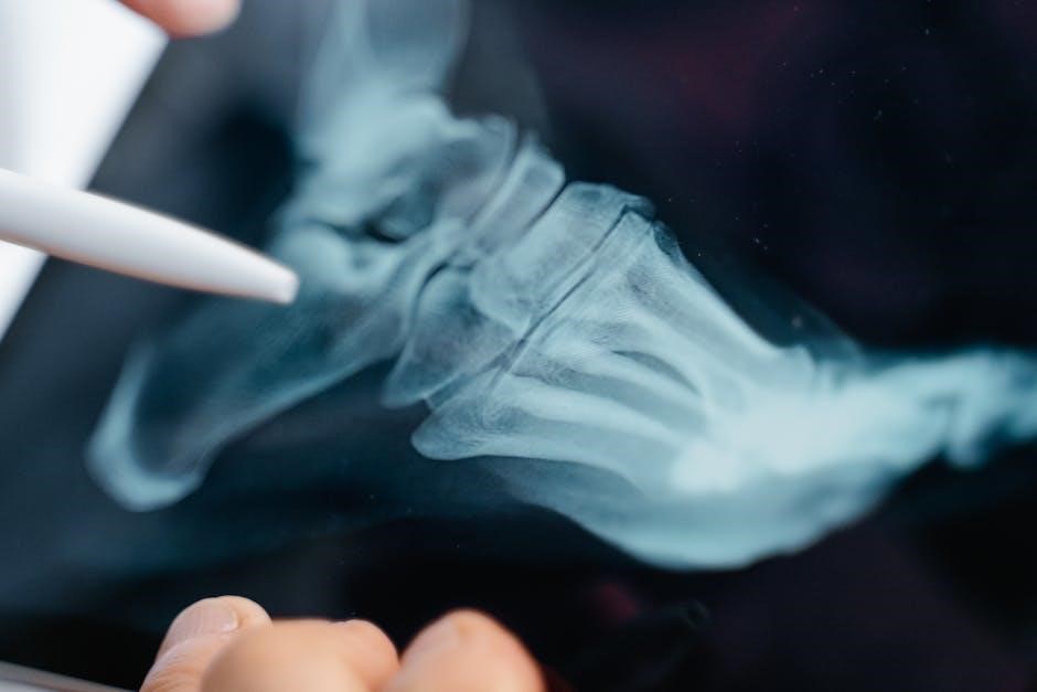
9.1 Role of X-Rays and MRIs in Diagnosing Infections
X-rays are essential for detecting bone involvement in diabetic foot infections‚ while MRIs provide detailed images of soft tissue and deeper structures. These tools help identify osteomyelitis‚ abscesses‚ or gas in tissues‚ guiding timely interventions. Early detection through imaging prevents progression and supports effective treatment plans‚ aligning with IWGDF recommendations for comprehensive foot care.
9.2 Using Wound Classification Systems for Documentation
Wound classification systems are crucial for standardized documentation‚ ensuring consistency in tracking ulcer progression and treatment. These systems categorize ulcers based on depth‚ tissue involvement‚ and infection status‚ aiding healthcare providers in communication and management. By incorporating such systems‚ clinicians can align with IWGDF guidelines‚ enhancing documentation accuracy and facilitating effective care plans tailored to patient needs.
Management and Prevention Strategies
Effective management and prevention strategies for diabetic foot care include wound care‚ proper dressing techniques‚ and recommendations for appropriate footwear and orthotics to prevent complications and promote healing.
10.1 Wound Care and Dressing Techniques
Proper wound care involves debridement‚ applying appropriate dressings‚ and maintaining a moist environment to promote healing. Regular monitoring for infection and using antimicrobial agents are crucial. Healthcare providers should follow evidence-based guidelines‚ such as those from the IWGDF‚ to ensure effective treatment and documentation of wound progression‚ enhancing patient outcomes and preventing further complications in diabetic foot management.
10.2 Recommendations for Footwear and Orthotics
Proper footwear is essential for preventing diabetic foot complications. Patients should wear shoes with a proper fit‚ adequate cushioning‚ and support to offload pressure points. Orthotics can be used to redistribute pressure and accommodate deformities like bunions or hammertoes. Healthcare providers should recommend footwear that aligns with the IWGDF guidelines‚ ensuring protection and comfort to enhance mobility and prevent further issues in high-risk patients.
Guidelines and Recommendations
The International Working Group on the Diabetic Foot (IWGDF) provides evidence-based guidelines for diabetic foot care‚ emphasizing prevention‚ classification‚ and treatment strategies to improve patient outcomes globally.
11.1 International Working Group on the Diabetic Foot (IWGDF) Guidelines
The IWGDF guidelines offer comprehensive‚ evidence-based recommendations for managing diabetic foot disease. They cover prevention strategies‚ classification systems for ulcers‚ and treatment protocols. These guidelines emphasize the importance of early detection through regular foot examinations‚ proper wound care‚ and multidisciplinary approaches. By following IWGDF recommendations‚ healthcare providers can significantly reduce the risk of complications‚ including amputations‚ and improve patient outcomes worldwide.
11.2 Practical Steps for Healthcare Providers
Healthcare providers should conduct regular foot exams‚ utilizing tools like monofilaments to assess neuropathy and Doppler studies for vascular evaluation. Proper wound care and dressings are essential for healing. Recommending appropriate footwear and orthotics can prevent further complications. Educating patients on self-inspection and prevention strategies‚ aligned with IWGDF guidelines‚ ensures comprehensive care and promotes better patient outcomes in managing diabetic foot conditions effectively.

Patient Education and Awareness
Patient education is crucial for managing diabetic foot health. Teach self-inspection techniques‚ proper foot care‚ and the importance of maintaining a healthy lifestyle. Encourage early detection of issues to prevent complications.
12.1 Teaching Patients to Self-Inspect Their Feet
Teaching patients to self-inspect their feet is vital for early detection of potential issues. Emphasize examining soles‚ toes‚ and heels for ulcers‚ redness‚ or swelling. Patients should check for changes in foot shape or sensation and report any concerns promptly. Use visual aids and guides to help them identify abnormalities and encourage daily checks to maintain foot health effectively.
12.2 Promoting Healthy Lifestyle Choices for Foot Health
Promoting healthy lifestyle choices is crucial for foot health. Encourage patients to manage blood glucose levels‚ maintain a balanced diet‚ and stay hydrated. Regular physical activity improves circulation‚ while avoiding smoking and excessive alcohol reduces vascular risks. Emphasize the importance of proper footwear‚ clean feet‚ and moisturizing to prevent cracks. These practices‚ along with weight management‚ contribute to overall foot well-being and reduce complications in diabetic patients.
Regular diabetic foot examinations are vital for early detection and prevention of complications. Adhering to IWGDF guidelines ensures comprehensive care. Referral to specialists and consistent follow-up are essential for optimal management and preventing severe outcomes.
13.1 When to Refer to a Specialist
Referral to a specialist is necessary for high-risk patients with deep ulcers‚ poor circulation‚ or signs of infection. Severe neuropathy‚ deformities‚ or non-healing wounds also warrant specialist care. Early referral ensures timely intervention‚ such as advanced wound management or vascular surgery. Guidelines recommend consulting podiatrists‚ endocrinologists‚ or vascular surgeons for complex cases to prevent amputations and improve outcomes.
13.2 Follow-Up Care and Monitoring
Regular follow-up appointments are crucial to monitor healing progress and manage diabetic foot conditions. Patients should be educated on self-inspection techniques to identify early signs of complications. Adjustments to treatment plans and footwear recommendations are made based on the patient’s evolving needs. Monitoring for neuropathy and vascular changes is essential to prevent future issues‚ ensuring comprehensive care.